Cystic Acne: focus
20/09/2023
Ingrown toenails or Onychocryptosis: intervention and medication
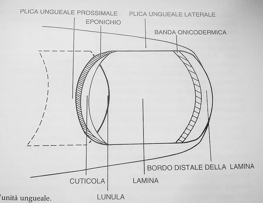
21/09/2023The nail is found at the ends of the fingers and toes. It is a poly-compound, keratinized lamina produced by specific cells. The set of structures that are involved in the formation and location of the nail plate is referred to as the nail unit which is composed of numerous specialized units, as shown in the image below.
The nail unit
The most important part of the nail unit is:
The germinal matrix (GM)
The germinal matrix is located below the proximal nail fold and is composed of keratinocytes that specialize in the production of the nail plate.
The matrix can be divided in two parts:
- distal, that produces the lower portion of the lamina
- proximal, that produces the superficial portion of the nail plate
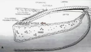
The distal portion of the lamina extends beyond the nail bed and above the digital sulcus and nail plica. Below, it extends the hyponychium to which connective fibers bind between the end of the phalangeal bone to the skin.
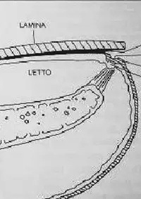
The lamina has a latero-lateral curvature that is more pronounced in the toenail.
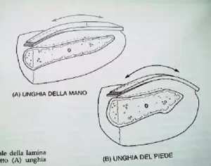
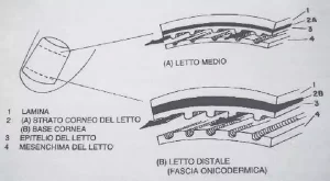
As the drawing of a nail unit cross-section shows, the stratum corneum component that lies between the epithelium of the nail bed and the lamina increases in the distal plate (B). Under normal conditions, this is evidenced by a decrease in pink coloration (transparency of blood vessels) between proximal and distal areas.
Nail physiology
The nail lamina is a multilayered sheet of cuneiform cells that are tightly adhered to each other. The main functions of the laminae of the fingernails are to grasp small or elusive objects. Meanwhile, in the toenails, the lamina help to counteract and dissipate the pressure forces that occur when walking.
Nail growth
Fingernails grow by about three millimeters per month, meaning that it takes about six months for the lamina to be completely replaced. Toenails take two to three times as long to grow as fingernails.


