The complex anatomy of the nail
21/09/2023
Tyloma: a very particular callus
21/09/2023Ingrown toenails are an increasingly common podiatric pathology, the causes of which include the use of footwear that is unsuitable for the shape of the foot and/or being overweight.
The consequences of being overweight as an infant are numerous and occur as the bones, joints and skin structures are still in their formative stages. When a child begins to take their first step, if they are overweight, an excess of panniculus adiposus within the phalanges of the toe push the dermis and epidermis to encompass the lateral edges of the nail plate. This pressure causes the nail to curve, making it appear similar to the shape of a roof tile.

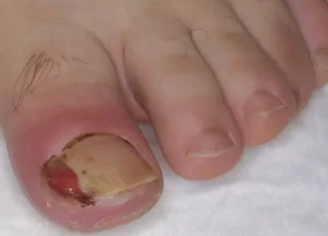
As an overweight child approaches puberty, their excess weight increases the pressure on the distal phalanges to the point where the nail will penetrate the lateral margin. This most commonly happens on the first toe. When the lateral margin of the nail plate breaks through the epidermis, contact occurs between the nail (lamina) and dermis and, therefore, between the nail and circulating immunocompetent cells. The body identifies the keratin that is cutting into the skin as a foreign body, which causes the area around the nail to become very inflamed. At this point, the passage of microorganisms (bacteria and/or fungi) spread into the dermis, triggering the release of immune defense cells such as neutrophil leukocytes. The ensuing ‘battle’ between the defending cells and the invading nail plate and microorganisms causes a considerable inflammatory reaction that can trigger the formation of reactive granuloma.
Reactive granulomas
A reactive granuloma is the result of an overgrowth of dermal tissue. They are delicate, can be filled with pus (neutrophils) and can bleed very easily. Reactive granulomas are extremely painful and the risk of superinfection is very high, meaning that prompt intervention is vital. Reactive granulomas are usually excised during surgery in order to remove the ingrown nail fragment. Ahead of surgery, the bacterial load of the infected area must be reduced in order to lower pain and to minimize the risk of infection.

Treating reactive granulomas
Reactive granulomas can be treated for two different reasons. They can be treated to reduce infection and pain before proceeding with a podiatric intervention to resolve both the granuloma and the ingrown nail. They may also be treated ahead of surgery for the purpose of reducing the bacterial load and decreasing the risk of infection during the operation.
Mistakes when treating reactive granulomas
Many methods used to treat reactive granulomas do not help in any way and can actually worsen inflammation. For example, the prescription and use of antibiotic creams are not recommended for two reasons. Firstly, the cream itself can cause occlusion and maceration of the granuloma, and secondly, the antibacterial properties of the cream do not get a chance to intervene as the lesion is open and is therefore frequently reinfected. Furthermore, the use of antiseptics such as iodopovidone or chlorine derivatives only irritate the lesion more, while warm water salt baths are even more deleterious.
Treating reactive granulomas
To dress reactive granulomas, first use Astringent Gel. A hydroalcoholic, Astringent Gel contains aluminum chloride hexahydrate and, as well as having antiseptic properties, can coagulate the proteins of the exposed cell to reduce the granuloma. The Gel vehicle allows the part to be treated without occluding. Apply Astringent Gel in the morning and evening. Do not cover with dressing. The photograph shows a reactive granuloma that has been ‘dried’ following Astringent Gel use.
Surgical intervention
The decision to intervene surgically is based on different factors, including:
- the failure of non-surgical treatments;
- intense pain;
- weakened plantar support (antalgic plantar support)
- patient’s need to solve the problem
Why avulsion is a mistake
Patients with ingrown toenails are, even today, unfortunately treated with the removal of the entire nail plate. This is a serious procedural error for many reasons:
- lamina detachment results in recurrence or worsening of ingrown toenails in most cases;
- removal causes the distortion of the distal phalanx;
- the postoperative period is painful;
- requires time-consuming dressings;
- there is a risk of infection;
- nail reconstruction is not guaranteed.
The following image shows the damage caused by lamina avulsion. The nail has regrown but it is severely dystrophic, the ingrown nail has reformed in the outer lateral margin, and the phalanx is deformed (babbitt phalanx).

Before removing the nail of the first toe, it should also be taken into account that this nail has considerable load-bearing functions while walking. In its absence, the soft parts of the toe and, sometimes, even the bone can become deformed and tip upwards. When the lamina regrows, its free edge impacts on the upturned margin of the toe and, as it cannot overcome the obstacle, it grows to become deformed. In these cases, a soft-part correction surgery must be performed which involves opening a groove at the base of the phalanx and sliding the raised portion of the phalanx downward before suturing.

Nail phenolization
The nail plate will only penetrate the lateral margins of the toe, where the plate naturally folds in order to slide into the nail groove. The removal of this curved, lateral portion of the nail while respecting the flat central area of the nail should be enough to overcome the issue. By removing the lateral portion without touching the nail matrix cells allows the regrowth and incubation of the lamina, but the question remains – how can this be done? In the past, destroying only the lateral portion of the nail matrix was often done with diathermic current coagulation, laser vaporization or through the application of cold liquid nitrogen. However, over time, these methods were abandoned as they posed real risks of damaging the periosteum and the underlying phalangeal bone, to which the nail matrix cells are very close. Today, the lateral portion of the nail matrix is removed with the chemical phenol.
Phenolization of the lateral portion of the nail matrix to treat ingrown nails
The standard treatment to overcome ingrown toenails by removing the lateral portion of the nail is phenolization surgery, which destroys lateral nail matrix cells through the use of phenol. Phenolization surgery has a high success rate and a low risk of side effects. It is a straightforward outpatient procedure and is performed in about thirty minutes with an added hour’s observation time. When discharged, the patient is able to walk painlessly.
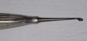
- A sharp spoon like the one in the illustration is used for excision.
- After ascertaining that anesthesia has occurred, the entire reactive granuloma is removed.
- A tightly tensioned tourniquet is applied.
- The superficial dermal infiltration is anesthetized starting at one centimeter above the lamina. Next to be anesthetized are the lateral side and the toe from the side that is to be operated upon.
- Local anesthesia should be administered using either carbocaine or analog without adrenaline. It is important not to use anesthesia with adrenaline as it can cause tissue necrosis through vasoconstriction. Although nerve trunk anesthesia can be performed, it is advisable to perform local anesthesia to avoid prolonged anesthetic effects or nerve damage.
- The toe to be operated on is sterilized with benzalkonium chloride solution. Do not sterilize with iodopovidone as it may interfere with phenol.
- The patient should be made to sit on the operating table, and the leg should be flexed so that the sole of the foot rests completely on the table top.
- Operate on only one toe per foot per session to limit phenol exposure.
- First visit: the patient will be clinically evaluated to determine their suitability for this type of surgery. Informed consent is achieved, Astringent Gel is prescribed for one week ahead of surgery, and patients are asked to come to surgery wearing either sandals or open-toed shoes as the first dressing may be bulky.
- Patient selection: there are no restrictions, although patients on antiplatelet and anticoagulant medications should be closely evaluated because of the risk of increased bleeding time after surgery.
- The lateral portion of the lamina is cut using a straight cutter similar to that in the picture.
- After the initial incision, a further three to four millimeters are cut by also incising the skin which will facilitate the removal of the nail that is usually lined by the skin.
- The cut portion of the lamina is hooked with a curved forcep such as the one shown. It is then lifted upward to disengage it from the area of the ingrown nail, before the subcutaneous portion of the lamina is detached with a firm, outward pull.
- If the operation is successful, a plow-shaped portion of the nail plate is removed where the handle is the outer portion that had become ingrown and the plow blade is the inner lateral portion.
- At this point, a cotton-coated wooden stick coated in liquid phenol is then applied.
- Liquid phenol at 85% in water is used (phenol in low concentrations is not effective)
- The applicator is inserted deeply into the incision and held in place for thirty seconds.
- Any smears of phenol are wiped off, before a second phenol application of thirty seconds is made thirty seconds after the first.
- The operation is completed and the tourniquet removed.
- The nail bed should be dressed with gauze and a band-aid will stop any bleeding.
- The patient should then be placed on a couch with the leg in an elevated position for 30 to 60 minutes, or the time required for a stable clot to form and for the patient to be able to walk.
- After making sure that bleeding has ceased, a layer of gauze medicated with PEG Ointment is applied and secured with a round of tape dressing.
- The first dressing should be maintained until the following day.
- Subsequent dressings of cotton gauze and PEG Ointment should be applied morning and evening until the crusting is detached.
- During this period, the operated foot should not be washed and must not get wet to prevent any liquid from entering the surgery crevice.
- Instead, to clean the foot, use an aqueous disinfectant, such as benzalkonium chloride, which can be poured onto a cotton pad then wrung out to remove excess disinfectant.
- Upon being discharged, the patient can walk and continue their day-to-day activities, but must suspend all sport for fifteen days.
- It is also advisable not to drive after surgery because of possible lack of sensation in the operated foot.


