Ingrown toenails or Onychocryptosis: intervention and medication
21/09/2023
Remedies for cracked heels
21/09/2023Tyloma is a term derived from Greek, which literally means “to harden”. This term should replace callus, Clavus, Corno, terms that should no longer be used because they do not define a specific clinical entity.
The clinical definition of Tyloma is: a small rounded area of hyperkeratosis at a plantar or dorsal rubbing point.
The hallmark of tyloma is hyperkeratosis.
Hyperkeratosis is the pathological thickening of the stratum corneum which can occur for a variety of reasons.

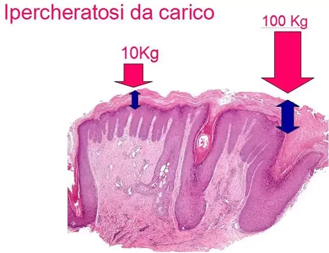
In the case of tylomas, the stratum corneum thickens and becomes hard (hyperkeratotic) as a result of pressure or friction. Vital epidermal cells act to defend and protect themselves against these mechanical pressures which further accentuates the thickness of the stratum corneum.


Plantar tylomas usually appear at the top of the metatarsals. This is especially true if the metatarsals are being pushed down due to a restriction in the tendons due to: hammer toe or loss of transverse arching or osteoarticular changes.
Dorsal tylomas form in areas where the foot rubs against the shoe, often above the interphalangeal joints or on the external side of the fifth toe.
Heloma is the name given to the hyperkeratotic lesions that form between the interdigital spaces of the toes. Helomas have different pathogenesis and treatment from tylomas.

Tyloma or wart?
Tylomas should be differentiated from other forms of hyperkeratosis on the feet and must be distinguished from plantar and dorsal warts.
Warts are caused by human papillomavirus (HPV) infection. This virus induces benign tumor growth characterized by marked hyperkeratosis meaning that lesions may be mistaken for tylomas, especially when they occur on the soles of the foot.
Differentiating a plantar wart from a tyloma is not always an easy task, which is why warts are often treated as though they were a tyloma and vice versa.
Warts vs. tylomas
| Wart | Tyloma | |
| Hyperkeratosis | Yes | Yes |
| Painful under pressure | Yes | Yes |
| HPV | Positive | Negative |
| Location | Anywhere | Pressure points |
The only way to ascertain whether a lesion is a wart or a tyloma is to remove the hyperkeratosis with a gouge, preferably mounted on a micromotor.
After removal of this superficial layer of hyperkeratosis, it will be clear if there is a wart if the tissue underneath is soft and bleeds easily. Tylomas, on the other hand, are hard even when the first layer of skin is removed.
Tylomas vs. callous plaques
Tylomas are defined as localized hyperkeratosis. Instead, diffuse hyperkeratosis or callous plaques, are defined as a thickening of the stratum corneum that covers an extensive plantar area.
If the entire stratum corneum of the foot’s sole is covered in hyperkeratosis, this is a case of plantar tylosis.
Tylomas and pain
When pressure is applied to the area (e.g. through walking), tylomas cause intense pain, akin to that of having a sharp stone in the shoe or having a pin inserted into the skin. The pain is generally worse in the evening or after a walk, but can be provoked by putting any pressure on the tyloma. Warts that occur on pressure points can be equally painful, so the presence of pain is not a useful indicator in determining whether a lesion is a wart or a tyloma. Tylomas cause pain because the hyperkeratosis on the pressure point compresses the dermis and the nerve endings present below. The pain caused by the tyloma prompts the patient not to want to put pressure on the area where the tyloma occurs, which will alter their gait and cause other joints to become overloaded, inflamed and painful. Tylomas are not innervated or vascularized, so the term ‘neurovascular tylomas’ that can often be found in podiatry texts, should not be used. The thickness of a tyloma is usually more than one centimeter, as can be demonstrated when the tyloma is extracted with a biopsy punch. This is an intervention that occurs only in certain tyloma cases. Looking at the tyloma under histological examination, there is a clear alteration of the epidermis, which appears to be considerably thickened (acanthotic). In addition, the corneal lamellae that form the hyperkeratosis are stacked very compactly.
Examining the histology in more detail, it is clear that, in addition to the thickening of the epidermis, the dermal papillae that have an arteriole, push up to just below the the transition between the corneal lamellae of hyperkeratosis and the cells of the epidermis.
This explains why the tyloma bleeds very easily when it is decapitated by cutting too deeply.

Plantar warts have a histologic appearance very similar to that of tylomas, except the cells of the epidermis that are infected with HPV are easily spotted upon high magnification.
Why do tylomas form?
When tylomas appear, it is a sign that something has changed in the way the foot provides support, or that something isn’t working properly while walking. It’s important to investigate the exact cause of why the tyloma has formed, as these could be patient-related, such as when there has been an increase in body weight causing more pressure on the foot. Similarly, tylomas could form after weight loss with reabsorption of the plantar panniculus adiposus. In other cases, there may have been muscle-articular damage that has forced the patient to modify the way in which they walk. Inflammatory or arthritic phenomena affecting the bones and joints of the feet with modification of bone and joint profiles are also causes of tyloma. Among foot-related causes, the most frequent is musculotendinous alteration leading to hammer toe and the consequent lowering of the metatarsal heads.
Sometimes, the causes of tylomas can be traced back to footwear, particularly the use of safety shoes or wooden clogs. They can also appear after recreational activities such as very long walks with uncomfortable footwear on rough terrain, etc.
What are the consequences of tylomas?
Regardless of the cause of tylomas, they cause pain every time they are exposed to pressure. To avoid feeling this pain, the patient may avoid leaning on the part of the foot where the tyloma is present and in doing so, alters the supports of the foot and their gait (antalgic walking). The consequence of this is that other osteo-muscle-tendon structures are overloaded and can become inflamed and painful, with the knees and hips the areas most likely to be affected.To avoid this, it is crucial that the tyloma be treated as soon as possible.
Investigations into tylomas
It is usually sufficient to simply observe the foot in order to locate the support and nonsupport points, and to assess any postural defects.
However, barometric footplates may also be used in order to fully understand the foot. These involve having the feet placed upright on a piezoelectric matrix insole that is capable of translating any vertical pressures exerted onto the insole into electrical signals. Through a special program, these signals are converted into light signals that form a two-dimensional multi-color map. In the map, blue represents an area of minimum pressure, while red shows maximum pressure.

While active matrix barometric treadmills are able to give measurements when the patient is more static, insoles can be inserted directly into the patient’s shoe while in the clinic, allowing real time gait analysis images to be transmitted to the computer. Gait analysis and measurement allows for a more complex understanding of displacement loads while walking.
These, and other available measurements, are able to identify and document any overloads and to determine precisely where and how to counter this with the construction of appropriate footplates.

Treatments for tylomas
As tylomas are hyperkeratotic defense reactions, treatment should aim to restore proper plantar support or the correct positioning of the toes. Unfortunately, it is not always possible to act on the causes alone and, instead, tylomas must be treated more directly to reduce or eliminate the pain.
There are a wide range of factors to be considered about the patient before any treatment can take place.
Conditions to be considered before selecting the tyloma treatment course are:
- Age
- Work activity/leisure habits
- Weight, specifically overweight or obesity
- Concomitant diseases: diabetes, arthritis, psoriasis, gout, etc.
- Osteoarticular history, such as fractures or prostheses
This information is used to decide which course of action to take in the treatment of Tiloma.
After clinical inspection, gait observation and barometric treadmill studies or gait analysis, a suitable treatment can be decided upon.
Treating tylomas
Interventions can vary and can be:
- Provisional, or aimed at returning to pain-free walking in the immediate term
- Complementary and provisional, with the aim to control the hyperkeratotic reaction
- Permanent, aimed at correcting the causes that generated the tyloma
Provisional interventions
- Manual gouge pickling
- Motorized gouge pickling
Provisional Complementary Interventions
- Felting
- Corrective insoles
- Keratolysis
Long-term treatments
- Discharge insole
- Osteotendinous interventions
Pickling
The purpose of pickling is to reduce the thickness of the hyperkeratosis so that the pressure does not build up on the nerve endings of the dermis and thus cause pain.
Pickling can be carried out manually with a gouge or mechanically with a gouge mounted on a micromotor.
Manual pickling with gouge
The gouge is chosen according to the maximum diameter of the Tiloma to be pickled. The rule is: choose a gouge that is one or two millimetres narrower than the diameter of the Tiloma.
Hold the handpiece tightly and exert tangential pressure so as to remove a thin curl of stratum corneum with each pass.
Stop when the hyperkeratosis of the Tiloma is a few millimetres below the support plane.
Forcing the removal may lead to amputation of a dermal papilla with consequent pain and bleeding.

Mechanical pickling with a gouge mounted on a micromotor
The gouge can be mounted on a handpiece with linear movement, which in turn is mounted on a micromotor. In this case, the gouge mechanically performs a forward/backward traverse of about 2 mm and the traverse frequency can be adjusted by the speed of the micromotor.
In this system, it is the gouge that performs the removal of the stratum corneum curl without having to apply muscle force.
Mechanical pickling with a gouge mounted on a micromotor is more sensitive and professional than manual pickling.
Provisional Complementary Interventions
Felting
Felting is a simple and effective way to remove pressure from the area.
A temporary measure, it involves the use of felt, which is a non-woven cloth made of the fur of various animals (often sheep’s wool), that is then compacted and felted.
As an alternative, rubber latex foam (synthetic foam is not recommended) may be used. Felt can be of different thicknesses and with various resistance to pressure, and may be cut to its full thickness to provide complete pressure relief. The tyloma should be undergoing pickling treatment and the felt provides protection during this time. The area to be relieved of pressure is identified, before the partially-hollowed out piece of felt is applied. The felt piece is applied to the skin by the side covered in glue which allows it to adhere to the skin.
To ensure the stability of the felting, however, apply a perforated plaster (Mefix) above and on the skin all around.
This dressing can last for a maximum of one week before being changed.
Felting has two main drawbacks: when placed in the plantar area, it raises the foot, which can throw foot support and balance off kilter. Felting also prevents the covered area from being cleaned while the dressing is in place.
Corrective insoles
Plantar insoles with a thickness of about 5 mm can be used to reduce pressure. The insole material can be natural (e.g. latex or cork), or synthetic (expanded resin, silicone, etc.).
The perimeter of the area to be relieved of pressure is traced with a demographic pen on the individual’s foot. By positioning the insole on the sole of the foot, the imprint of the pressure zones is obtained directly into the insole. The edges are then cut and smoothed, and the insole is inserted into the shoe.
Insoles are also a temporary solution. Depending on their composition, after the initial period, the insole tends to compress which cancels out any relief it previously gave.
Keratolysis
Keratolytic agents induce the detachment of the corneocytes from the superficial stratum corneum. In turn, this reduces both the adhesion of the corneocytes and the thickness of the stratum corneum. The most commonly used keratolytic agents are salicylic acid and glycolic acid, with the former being particularly well-known.
There are various, commercially-available topical products with keratolytic agents, however these are usually for the treatment of warts and are not suitable for tylomas.
Moreover, these creams are often packed with various ingredients.
Although there are some lotions with salicylic acid for tyloma treatment, the concentrations of the acid are not adequate enough to ensure tyloma removal.
Indeed for optimal keratolysis, salicylic acid must be used at a concentration of 30%.
A pharmacist will be able to prepare treatment with 30% salicylic acid in flexible collodion. The formula is as follows:
Keratolysis with salicylic acid in flexible collodion
- 30% salicylic acid in flexible collodion
- 3g salicylic acid
- 5g, 5% flexibile collodion
- 1ml ethyl ether
- 1ml ethyl alcohol
Label marked: ‘external use only’
Store the solution in the refrigerator to prevent it from drying out.
Flexible collodion as a carrier allows for the easy and precise application to the point of hyperkeratosis.
Upon contact with air after application, elastic collodion transforms from a liquid into a solid. This film adheres to the skin and brings the concentrated salicylic acid into contact with the stratum corneum. There is no need to cover or medicate.
The use of the keratolytic is recommended between one pickling treatment and another, to maintain the healthy depth of the stratum corneum, or before a pickling to make it easier or after it to maintain the result obtained.
Keratolysis with cream
To perform an effective keratolysis, the cream used must have a sufficient quantity of exfoliating acids in order to thin the stratum corneum and decrease pain.
The keratolytic cream (Glycocream +) proposed by DermaClub is composed of three associated acids: salicylic acid, glycolic acid and succinic acid. The combination of these acids at a concentration that is safe for skin, induces effective desquamation that can treat tylomas. .
Long-term treatments
The most common, long-term treatment for tylomas is the creation of an insole (for information on the theory and practice of a relief insole, refer to specific texts). A custom-made insole is prepared to prevent the foot from putting too much pressure on the area where the tyloma has formed. As soon as this area no longer experiences excessive weight or pressure, the tyloma will be spontaneously reabsorbed.
However, it may not always be possible to create an insole due to the particular anatomical conditions of the foot. In such cases, orthopedic surgery is often necessary.
Osteotendinous interventions
The osteoarticular and tendon deformities that are the root causes of tyloma can be treated by an orthopedic surgeon. Bone segments and tendons can be lengthened, shortened or lifted in order to restore the correct support for the foot, thus avoiding the formation of tyloma in the future.
In practice, surgery options will vary greatly depending on factors such as age and the general health of the patient.
For further information on the subject, please refer to more specific papers.


